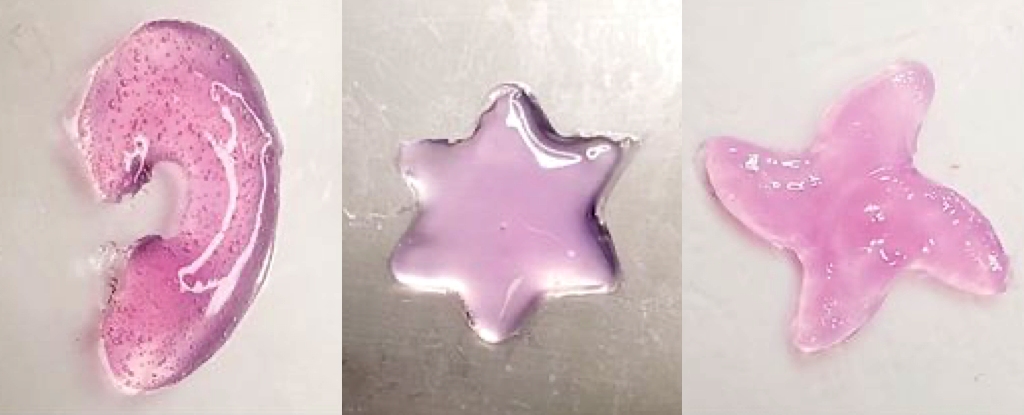
In a advancement, scientists at the California Institute of Technology (Caltech) have developed a revolutionary method called deep tissue in vivo sound printing (DISP), enabling the 3D printing of materials directly inside the body using ultrasound. This innovative technique, already demonstrating promising results in animal trials, opens up exciting new possibilities for targeted drug delivery, advanced tissue repair, and the creation of implantable sensors.
The Innovative DISP Method: Precision Printing with Sound
The DISP method hinges on the injection of a specialized bioink into the body. This bioink’s composition is carefully tailored to its intended function, but it invariably contains polymer chains and crosslinking agents, both essential for the formation of a hydrogel structure. To prevent premature hydrogel formation, the crosslinking agents are ingeniously encapsulated within lipid-based particles known as liposomes. The outer shells of these liposomes are engineered to break down when exposed to a temperature of 41.7 °C (107.1 °F), a temperature only slightly above normal body temperature.
The 3D printing process is initiated using a focused ultrasound beam. This beam precisely heats the targeted area, causing the liposomes to rupture and release the crosslinking agents. Consequently, a hydrogel forms exactly where desired within the body. Unlike earlier methods that relied on infrared light, which has limited penetration depth, ultrasound can reach much deeper tissues, including muscles and organs. This capability dramatically expands the potential applications of in-body 3D printing.
Researchers can create intricate 3D shapes, such as stars and teardrops, with remarkable precision by meticulously controlling the ultrasound beam. This level of control is paramount for various biomedical applications, ensuring that the printed material perfectly conforms to the specific requirements of the targeted tissue or organ.
Versatile Applications: Transforming Medicine
Animal tests have showcased the versatility of the DISP method across several crucial areas:
- Tissue Repair and Replacement: The hydrogel can serve as a scaffold for artificial tissue, replacing or repairing damaged tissue. In rabbit trials, researchers successfully printed artificial tissue at depths of up to 4 centimeters (approximately 1.6 inches) below the skin. This suggests that DISP could accelerate the healing of wounds and injuries, particularly if cells are incorporated into the bioink.
- Targeted Drug Delivery: The bioink can be loaded with therapeutic drugs for precise and localized delivery. In a study involving 3D cell cultures of bladder cancer, the chemotherapy drug doxorubicin was incorporated into the bioink. Hardening the bioink into a hydrogel using the DISP method resulted in a slow, sustained release of the drug over several days, leading to significantly more cancer cell death compared to traditional injection methods.
- Implantable Sensors: By adding conductive materials such as carbon nanotubes and silver nanowires to the bioink, researchers have created bioinks suitable for implantable sensors. These sensors can monitor a range of physiological parameters, including temperature and electrical signals from the heart or muscles, opening up possibilities for continuous monitoring of patients with chronic conditions or those recovering from surgery.
Implications and Future Directions: A New Era of Personalized Medicine
The DISP method represents a significant leap forward in biomedical engineering, offering a minimally invasive approach to creating complex structures and delivering therapies directly within the body. The ability to 3D print materials deep within tissues and organs has profound implications for personalized medicine and the treatment of various diseases and injuries.
A key advantage of DISP is its biocompatibility. According to the researchers, no toxicity from the hydrogel was detected in animal tests, and any remaining liquid bioink is naturally eliminated from the body within seven days. This is crucial for ensuring the safety of the method for future clinical applications.
While the results from animal studies are highly encouraging, further research is essential to translate this technology to human applications. The next steps involve testing the method in larger animal models and, ultimately, conducting clinical trials in humans.
Looking ahead, the researchers envision integrating artificial intelligence (AI) into the DISP system. This could enable autonomous and highly precise printing within moving organs, such as a beating heart, revolutionizing the treatment of cardiac conditions and other dynamic physiological processes.
The publication of this research in the journal Science underscores the significance of this breakthrough. The DISP method holds immense potential for transforming our approach to tissue engineering, drug delivery, and medical device implantation, ultimately leading to improved patient outcomes and a better quality of life.
Original Source


