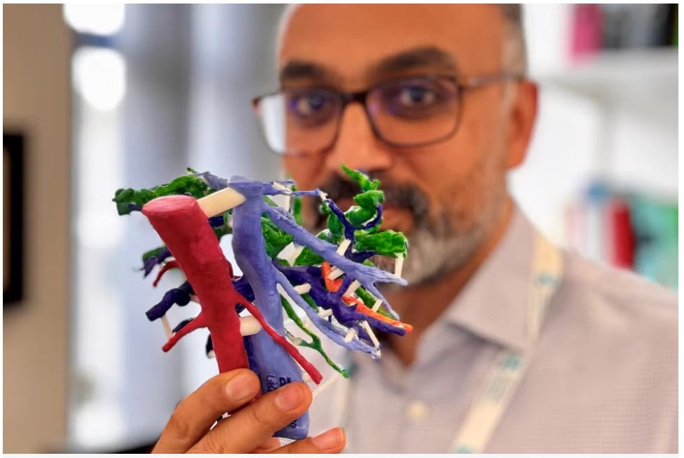
Surgeons at University Hospital Southampton have introduced an innovative approach to complex cancer surgery by utilizing 3D printed models of patients’ liver. This groundbreaking technique, supported by the Planets Cancer Charity, involves the creation of customized models based on CT and MRI scans of individuals diagnosed with hilar cholangiocarcinoma, a form of bile duct cancer.
The initiative addresses the need for precise surgical planning and aims to optimize outcomes by allowing surgeons to evaluate the feasibility of tumor removal before undertaking irreversible steps during the operation. Historically, the decision on whether a tumor could be safely removed was deferred until the surgery was in progress. However, the integration of 3D printing technology now provides a tangible solution to this longstanding challenge.
Consultant hepatobiliary and pancreatic cancer surgeon, Arjun Takhar, underscores the increasing role of 3D printed models in pre-operative decision-making. These models facilitate a comprehensive understanding of the anatomical relationships between tumors and organ structures, offering invaluable insights that were previously unavailable until the actual surgery. Mr. Takhar emphasizes that in the case of hilar cholangiocarcinoma, surgeons often face uncertainty until the last minute regarding the feasibility of complete tumor removal.
The 3D printing process involves the analysis of CT and MRI scan data, allowing for the creation of highly detailed physical replicas of patients’ livers. Surgeons can then use these models to assess whether a tumor can be safely removed, a critical determination that was previously deferred until the surgical procedure was underway. This proactive approach reduces the likelihood of irreversible steps being taken before confirming the operability of the tumor, thereby enhancing both short-term and long-term outcomes for patients.
The Planets Cancer Charity, which supports patients with various cancers, including pancreatic, liver, colorectal, abdominal, and neuroendocrine cancers, played a pivotal role in funding this initiative. The charity received a £2,000 grant from The Hospital Saturday Fund to support the pilot project. The use of 3D printed models not only aids in surgical decision-making but also presents an opportunity to educate and train future surgeons. The technology offers a unique advantage in teaching trainee surgeons the intricacies of liver anatomy in relation to complex tumors, further contributing to advancements in medical education.
The pilot project aims to assess the effectiveness of using 3D models in determining operability without the need for invasive procedures. Additionally, it seeks to evaluate whether assessing operability with a physical model surpasses the traditional method of relying solely on CT and MRI scans. The potential success of this initiative could pave the way for the adoption of 3D printing technology in the treatment of other liver tumors, marking a significant step forward in the integration of cutting-edge technology in the field of surgical oncology.
Subscribe to AM Chronicle Newsletter to stay connected: https://bit.ly/3fBZ1mP
Follow us on LinkedIn: https://bit.ly/3IjhrFq
Visit for more interesting content on additive manufacturing: https://amchronicle.com


