


At a rapidly growing rate, the medical industry continues to recognise the added value of additive manufacturing (AM or 3D Printing) for a vast range of medical applications. AM is now used in the development of new surgical cutting and drill guides, orthopedic implants, and prosthetics as well as the creation of patient-specific replicas of bones, organs, and blood vessels. Medical uses for 3D printing also include creation of anatomical models, tissue and organ fabrication and pharmaceutical research regarding drug dosage forms, delivery, and discovery.
Benefits of 3D printing in surgery
The application of 3D printing in medicine can offer many benefits, including: the customisation and personalisation of medical products, drugs, and equipment; cost-effectiveness; increased productivity; the democratisation of design and manufacturing; and enhanced collaboration. With the use of 3D printing, one can create patient-specific organ replicas so that surgeons can be use to practice on before performing complicated operations. This technique has been proven to speed up procedures and minimise trauma for patients. It provides easy and better diagnostics and understanding to surgeons. For instance, size of heart valve varies from person to person and with the availability of this technique, it is easy to plan exactly the sizing. India is at par with rest of the world in terms of medical advancement. In a country like India where there is scarcity of organs donation this technique is quite helpful and evolving. Around 20 cases per year end up with the use of this technology in any country, same like India.
“3D printing is a compelling new technology that has the potential to revolutionise cardiac intervention,” says Dr Ajay Kaul, Chairman and HOD, Cardiothoracic and Vascular Surgery, BLK Heart Centre.
Not many cases end up employing the use of 3D, but till now around 15-20 cases have used this technology across India out of which six are from BLK Heart Centre. It is definitely going to improve in India. When we started working upon it since last year, it was difficult for us to coordinate with the printing and its technology but now we are completely able to diagnose and feel easy to use. Similarly many doctors are unaware of this, but very soon they will come across and will improve.
Define 3D Printing
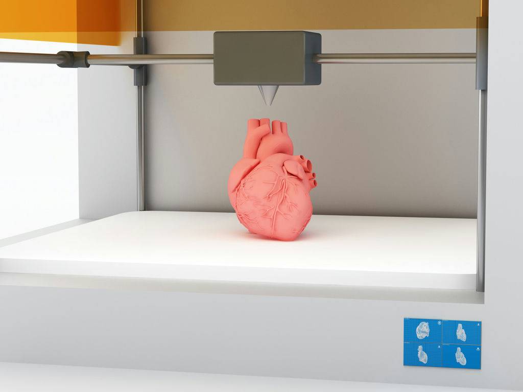
Three-dimensional (3D) printing, also known as additive manufacturing, is a manufacturing method in which involves taking a digital model or blueprint of the subject that is then printed in successive layers of an appropriate material to create a new version of the subject. The 3D printing process uses materials—such as plastic, metal, ceramics, powders, liquids, or even living cells—in layers to produce a 3D object. 3D printing is expected to revolutionise medicine and other fields.
There are about more than twenty 3D printing processes, which use varying printer technologies, speeds, and resolutions, and hundreds of materials. These technologies can build a 3D object in almost any shape imaginable as defined in a computer-aided design (CAD) file. (In a basic setup, the 3D printer first follows the instructions in the CAD file to build the foundation for the object, moving the print head along the x–y plane. The printer then continues to follow the instructions, moving the print head along the z-axis to build the object vertically layer by layer. It is important to note that two-dimensional (2D) radiographic images, such as x-rays, magnetic resonance imaging (MRI), or computerised tomography (CT) scans, can be converted to digital 3D print files, allowing the creation of complex, customised anatomical and medical structures.
3D Printing in the field of structural heart diseases
3D printing is a powerful technology with the potential to significantly change the practice of medicine. In the field of structural heart disease, this rapidly evolving technology can make a powerful impact. Current conventional cardiac imaging modalities such as echocardiography (EKG), cardiac computed tomography (CT) or magnetic resonance imaging (MRI) primarily utilise two-dimensional (2D) methods that require significant expertise and experience to interpret. In the field of paediatric or congenital cardiology, complex structural heart disease requires precise anatomical delineation before intervention. Consider a heart no larger than a walnut with multiple levels of abnormal connections.
3D printing produces a replica of the patient’s anatomy. In patients with complex congenital heart disease (CHD), this allows precise understanding of the patient’s anatomy and the resultant physiology. This technology solves some of the challenges of 2D imaging by enabling more informed decisions and precise pre-surgical planning. For surgical planning, 3D models enable detailed planning based on a physical model that can be held, manipulated in real 3D space, and scrutinised to plan various aspects of the surgery, including surgical approach, incision, cannulation technique, etc.
This sort of precise pre-surgical planning may lead to shorter operative times and fewer operative complications. Shorter cardiopulmonary bypass time, circulatory arrest time and fewer residual lesions requiring re-intervention are desired outcomes of precise pre-surgical planning from 3D printed models. These improvements in the operating room may translate to quicker recovery and a shorter post-operative hospital stay. Cardiac 3D printing is a nascent field, and at this time there is limited data to prove these beneficial outcomes. However, utilisation of this technology is growing, and, with time, the potential benefits of 3D printing will evolve drastically.
Diversity in 3D printing
With applications in congenital heart disease, coronary artery disease, and surgical and catheter-based structural disease, 3D printing is a new tool that is challenging how we image, plan, and carries out cardiovascular interventions. 3D printing has made a huge difference in the field of surgery of heart and aneurysm. Usually this method is popular in planning of heart operation, spine and other orthopaedic procedure. As this has nothing to do with the patient’s body during surgery there are no ill effects on them. Any organ or tissue can be made in nearly any imaginable geometry through the translation of x-ray, MRI, or CT scans into digital. In this way, 3D printing has been used successfully in the healthcare sector to make both standard and complex customised prosthetic limbs and surgical implants, sometimes within hours.
In the upcoming days, this itself will grow and develop with more advanced 3D models along with functional models where materials are more like the organs. Functional models along with organ like material could help us understand the diagnosis much better.
Advantages over traditional method
Therapies based on tissue engineering and regenerative medicine are being pursued as a potential solution for the organ donor shortage. The traditional tissue engineering strategy is to isolate stem cells from small tissue samples, mix them with growth factors, multiply them in the laboratory, and seed the cells onto scaffolds that direct cell proliferation and differentiation into functioning tissues. 3D bio printing offers additional important advantages beyond this traditional regenerative method.
Source: www.expressbpd.com


