


Out of all the unique abilities that helped us humans to win the evolutionary race, our ability to manufacture tools and heal ourselves has been instrumental in the story of our species. 3D BioPrinting combines these very two things that have shaped our civilization.
The ability to fabricate complex 3-dimensional structures that can house and nurture different kinds of cells will not only give us the ability to fabricate and replace essential parts of our bodies but open up an entire plethora of solutions, models, and applications which will undoubtedly change the face of modern medicine and life sciences.
Life sciences are the study of living things, living things are made up of cells organized into tissues organized into organs, and finally organisms. It has been a century since the first animal cells were isolated and grown outside a body and the time has finally come when scientists are studying and replicating the next level of biology in the form of tissue engineering.
Over the short span of its existence, Bioprinting has become the primary tool for tissue engineering to replicate the 3D organizational architecture that tissues possess naturally.
Cell culturing is the process of growing cells outside a biological system in a controlled and sterile environment. Celling culturing is limited by a property of cells called “contact inhibition” which causes the cells to stop growing in close proximity to each other for more than a few layers. Cells also cannot perform most of their unique functions until they are arranged and surrounded by other types of cells mimicking tissue architectures.
Understanding and replicating biological systems through 2D cell culturing has limited potential which has been explored and exhausted in the opinion of many scientists. This is where 3D bioprinting comes in to save the day, layering cell-laden biocompatible materials to form tissues and organoids that mimic in-vivo conditions for cells, allowing them to proliferate and form networks of communication with each other realizing their true nature.
How Does a BioPrinter Function?
Bioprinters
Many institutions and companies have realized the potential of this technology and have been funneling resources into developing it further.
A bioprinter as its name suggests prints biocompatible materials using a needle. Similar to a regular printer that prints ink onto paper, a bioprinter prints with bio-ink, which is a mix of living cells and a material that holds the cells and provides nutrients for the cells to grow. These droplets are carefully placed layer by layer to create a 3D structure that mimics the structure of living tissues or organs.

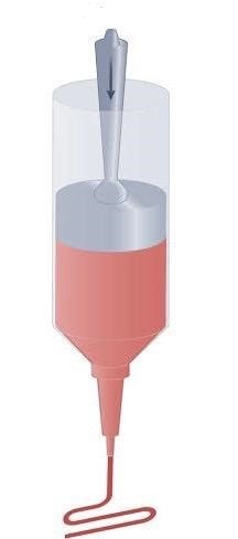
pushes the bioink through the nozzle on a
sterile surface according to a pattern the user wants.
The printer uses a computer-aided design (CAD) program to create a 3D model of the tissue or organ that needs to be printed. The model is then translated into instructions that the bioprinter uses to create the desired structure. The 3D structure is converted to Gcode which the printer can read. The g code functions similarly to binary code for computers.
All Bioprinting processes are performed with Bio-compatible or cell-friendly materials that are referred to as BioInks that can mimic the properties of matrices in natural tissues. The bioink contains cells suspended in a viscous fluid which provides nutrients for a cell’s growth and a structure to grow into. The composition of the bioink can vary depending on the type of tissue or organ being printed. For example, bioink used for printing bone tissue may contain calcium phosphate, while bioink used for printing blood vessels may contain extracellular matrix proteins like collagen. The bioink must be carefully designed to support the growth and function of the living cells, ensuring that the final printed tissue or organ is viable and functional.
Thus the printer deposits the bioink in a precise pattern, layer by layer, extrusion. The printer nozzle releases the bio-ink in layers, which are then arranged to create the desired three-dimensional structure. Once the structure is complete, it can be placed in an incubator where the cells can grow and mature into living tissue.

3D bioprinting started with simple 2D structures like a simple 1-layer tissue and from that it came a long way when for the first time Organovo a US-based company printed a viable liver tissue in 2013. It was a massive breakthrough for bio 3D printing. Modern-day bioprinters can print complex 3D structures such as blood vessels and the cornea of the eye. Recently an Israeli scientist Tal Dvir printed a small-sized heart using FRESH printing technology. It does not beat and is not suitable for human use and size of a rabbit heart but is a milestone covered in the journey of printing working organs and promises great results for printing viable tissues for organs. The main problem researchers are concerned about now is vascularisation inside the organ which is an integral part of organ printing, as without vascularisation the printed organ is not viable.
The future prospects of 3D bioprinting are incredibly promising. Here are some potential areas of growth and application. One of the most exciting prospects for 3D bioprinting is the potential to create functional, transplantable organs. This could significantly reduce the waiting time and improve outcomes for patients in need of organ transplants. 3D bioprinting could enable the creation of personalized tissues and organs that match a patient’s specific needs, reducing the risk of rejection and improving treatment outcomes. 3D bioprinting could provide a powerful tool for modeling diseases and testing new therapies. Researchers could print 3D models of organs affected by the disease to understand better how diseases develop and how treatments can be optimized. 3D bioprinting could also be used to test new drugs and therapies in a more accurate and reliable way. By creating 3D models of organs, researchers can test how drugs interact with the tissues and organs in a more realistic environment. 3D bioprinting could have applications in veterinary medicine as well. For example, it could be used to create prosthetics or implants for animals or to print tissues for testing new veterinary drugs.
Overall, the future of 3D bioprinting is incredibly exciting, with the potential to transform medicine and improve the lives of millions of people. With continued research and development, we can expect to see even more impressive applications and breakthroughs in the future.
Kreator3D, a Chennai-based startup, developing and manufacturing 3D BioPrinters is the newest player in the field with their product “BioForm” in the beta testing phases, they are on track to make considerable strides in the BioPrinting space with plans to provide eco-system-like solutions that cater to Bio-Convergence research and development and are hoping to become a key catalyst in the revolution of Healthcare and Medicine.
Subscribe to AM Chronicle Newsletter to stay connected: https://bit.ly/3fBZ1mP
Follow us on LinkedIn: https://bit.ly/3IjhrFq
Visit for more interesting content on additive manufacturing: https://amchronicle.com


