[vc_row][vc_column][vc_column_text]“On thorough work up we diagnosed him to have secondary chondrosarcoma from sacral bone.”, said Dr. Suman. “Removal of sacral bone tumor is a challenging job because of its close relation to vital blood vessels, pelvic viscera and nerve roots which passes through the sacral bone, and adding to the above, these bone tumors require Excision with good margins. (around 2-3 cm healthy tissue )”
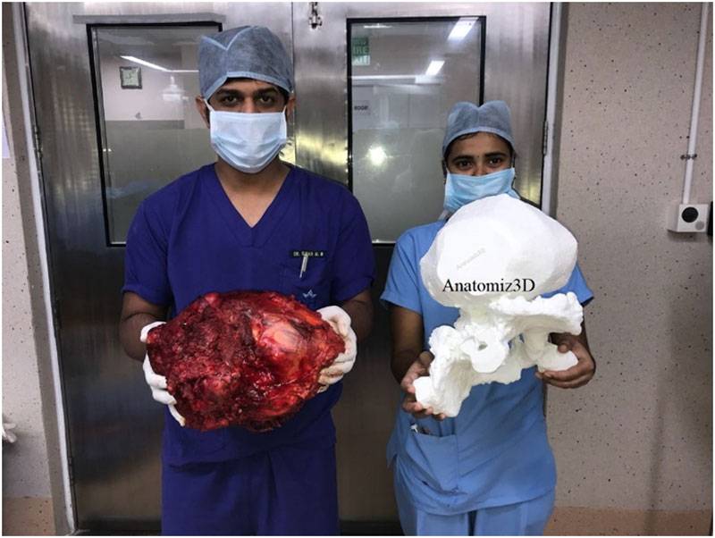
Anatomiz3D was contacted to provide a 3D Printed anatomical replica for this patient. When Dr.Suman contacted us to reconstruct an anatomical model for this case, we were quite astonished with the enormity of the condition. It was supremely important to ensure the accuracy between the margins of the bone and the tumour. The model was designed and 3D Printed over multiple builds comprising a total of 48 hours print time.
“We got a 3D reconstruction of patient pelvic and sacral bone. It helped us to plan our surgery precisely, excise tumor with adequate margins and avoid the complications which are associated with sacral tumor surgeries. “, Dr. Suman stated. “3D model is quite a handy tool which gives a realistic sized reconstruction of bone and tumor which helps in accurate planning and execution.”
The extent to which 3D Printing is showing its relevance in the clinical field is only growing every day. Anatomiz3D is excited to imagine the possibilities of the future.[/vc_column_text][/vc_column][/vc_row][vc_row][vc_column width=”1/3″][vc_single_image image=”3730″ img_size=”large” add_caption=”yes”][/vc_column][vc_column width=”1/3″][vc_single_image image=”3731″ img_size=”large” add_caption=”yes”][/vc_column][/vc_row]


